Researchers have mapped how neuronal connections develop in the healthy human fetus. Their results were published online on Wednesday in Science Translational Medicine.

Using functional magnetic resonance imaging, or fMRI, researchers at Wayne State University School of Medicine in Detroit studied the brains of 25 healthy second- and third-trimester fetuses. With fMRI, the team could detect neuronal circuits that connect one part of the brain to another. They saw that the neuronal connections that cross from one hemisphere of the brain to the other strengthen as a fetus ages. While those findings aren’t all that surprising, it’s interesting to think about what we could learn about the hidden development of fetuses. While many developmental disabilities such as autism and mental retardation have their start in the womb, they are usually diagnosed years after birth, which can make it difficult to understand their origin, say the researchers in the report. The team’s results show that fMRI techniques can elucidate the process of normal brain development and could help clinicians identify abnormal development earlier.
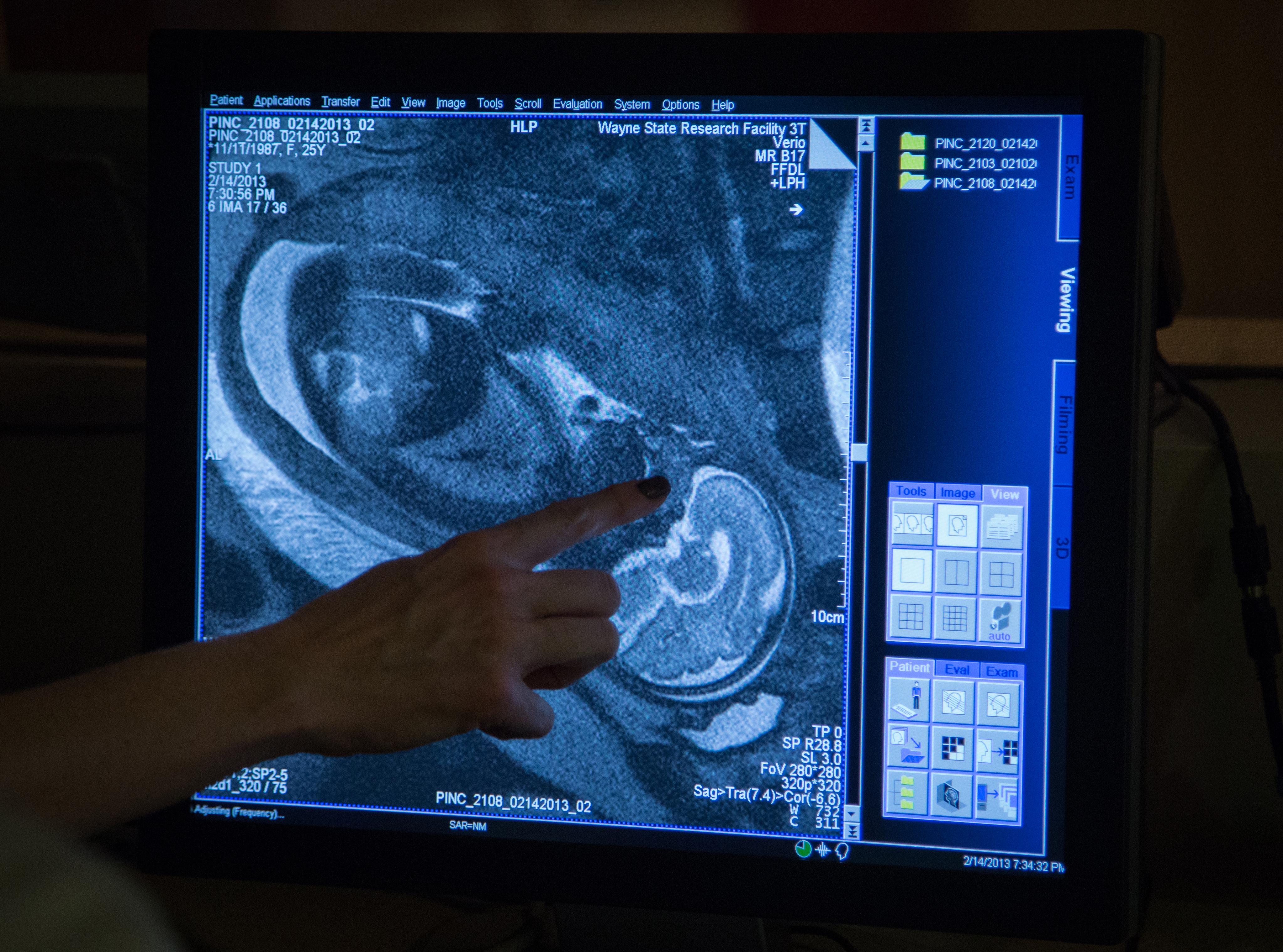
Image: A 31-week-old female fetus. Credit: Rob Widdis
Don’t settle for half the story.
Get paywall-free access to technology news for the here and now.
