Watching Live Cells
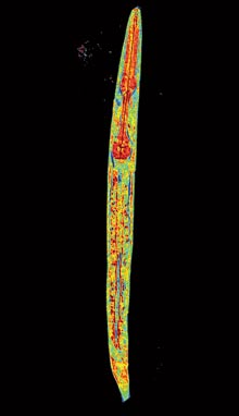
This is something you haven’t seen before: an image of a live, quarter-millimeter-long C. elegans worm whose internal organs, tail, and pharynx are clearly visible. The image was made by a microscope, under development at MIT, that renders cells and tiny organisms in 3‑D detail by exploiting the ways that different cell structures refract light and combining images made from several angles. In conventional microscopy, the cell or organism being studied must be treated with fixing agents and stains. “Our technique allows you to study cells in their native state with no preparation at all,” says Michael Feld, a professor of physics at MIT, who led the microscope’s development. In addition to imaging the worm, his group has imaged cervical-cancer cells, which could be seen shriveling up when treated with acetic acid. Because it can show how cells react to different compounds, the technology could be useful in drug screening.
Keep Reading
Most Popular
Large language models can do jaw-dropping things. But nobody knows exactly why.
And that's a problem. Figuring it out is one of the biggest scientific puzzles of our time and a crucial step towards controlling more powerful future models.
How scientists traced a mysterious covid case back to six toilets
When wastewater surveillance turns into a hunt for a single infected individual, the ethics get tricky.
The problem with plug-in hybrids? Their drivers.
Plug-in hybrids are often sold as a transition to EVs, but new data from Europe shows we’re still underestimating the emissions they produce.
Stay connected
Get the latest updates from
MIT Technology Review
Discover special offers, top stories, upcoming events, and more.