Early detection is key to surviving melanoma, a type of malignant tumor responsible for more than 70% of skin-cancer-related deaths worldwide, but “suspicious pigmented skin lesions” (SPLs) are so common it’s impractical for doctors to check them all out. Now MIT researchers have developed a tool that can analyze skin photos taken with a smartphone to determine which SPLs should be evaluated by a dermatologist.
The researchers, who include professors Martha Gray, SM ’81, PhD ’86, James Collins, and Regina Barzilay and postdoc Luis Soenksen, PhD ’20, made use of deep convolutional neural networks, machine-learning algorithms often used to classify images.

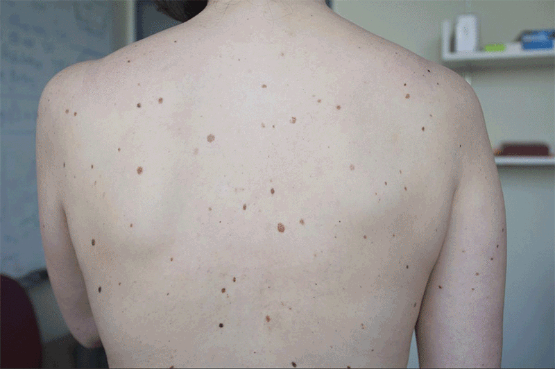
The team had dermatologists visually classify the lesions in 20,388 images from 133 patients at the Hospital Gregorio Marañón in Madrid and a number of publicly available images. The system was trained on 80% of those images and tested with the rest. It distinguished more than 90.3% of SPLs from nonsuspicious lesions, skin, and complex backgrounds. It also was able to classify the level of suspiciousness.
Don’t settle for half the story.
Get paywall-free access to technology news for the here and now.
