Brain images taken from hundreds of soldiers diagnosed with mild traumatic brain injuries suggest that methods for diagnosing concussions are inadequate in detecting damage. The results, part of the largest-ever imaging study of traumatic brain injury in the military, provide evidence that even brain injuries commonly classified as mild may lead to long-lasting damage.
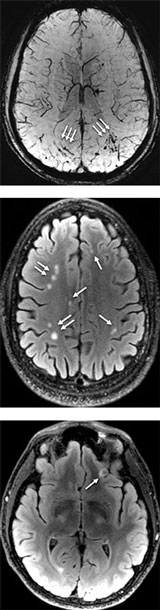

Researchers at Walter Reed National Military Medical Center observed abnormalities in the white matter—the part of the brain responsible for transmitting signals between different regions—of more than half the participants, most of whom had been diagnosed with at least one concussion. Gerard Riedy, a neuroradiologist at Walter Reed who led the research, says the large number of abnormalities seen in this study was surprising, and it undermines the conventional wisdom that a person with mild traumatic brain injury should have normal brain images.
More than 300,000 U.S. service members have been diagnosed with traumatic brain injury since 2000, often the result of blast-related trauma. Using imaging to detect damage could help doctors determine the most appropriate treatment. Often in concussion cases neither a CT scan nor an MRI reveals any signs of brain damage. And the clinical tools available for assessing the injury, which include a patient’s history, evaluations of cognitive skills like memory and attention, and tests of certain motor skills, require a large degree of subjective interpretation, says Riedy. Further, those assessments can be muddled by other conditions like post-traumatic stress disorder, which can cause many of the same symptoms.
Don’t settle for half the story.
Get paywall-free access to technology news for the here and now.
