Before scientists can build human organs in the lab, they need to figure out how to build tissues that work like those in the body. A new method, in which DNA acts like Velcro that makes cells stick to each other, could help pave the way toward building functional tissues that might one day comprise organs.
In nature, cells self-assemble into the complex three-dimensional architectures that comprise tissues. Biological function follows from this structure, and depends on the specific arrangement of cells, often of different types, in relation to one another. An individual cell’s behavior depends on signals from neighboring cells, and the collective behavior of the cells and tissues in an organ emerges from these 3-D relationships.
Advertisement
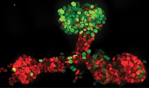
The cells in this microscope image were arranged in three dimensions using a new DNA-based technique. The cells stained green were meant to mimic cells that lead outgrowth during natural organ formation.
The new method employs DNA strands, attached to the outside of individual cells, to cause them to stick to surfaces—or other cells—that feature complementary strands, and assemble into prescribed arrangements. The researchers use it to programmatically build tissues, layer by layer.
This story is only available to subscribers.
Don’t settle for half the story.
Get paywall-free access to technology news for the here and now.
Other groups are taking a range of approaches toward building functional tissues (see “A Manufacturing Tool Builds 3-D Heart Tissue”). But compared to existing 3-D culture methods, the new one provides a greater level of control over “the ultimate tissue architecture,” argue its creators in a recent paper describing the research.
3-D printing of cells has become a popular way to arrange them for tissue engineering. But this method is hampered by the fact that it’s hard to keep cells alive and healthy throughout the printing process, and it can’t place cells with the precision that is needed, says Zev Gartner, a professor of pharmaceutical chemistry at the University of California, San Francisco, who led the research. Achieving “single cell resolution” is important, and the new method is able to do that, he says. Indeed, “high degrees of control and versatility” give this technique advantages over previously reported tissue assembly techniques, says Lisa Freed, a senior scientist at Draper Laboratory.
At this point the new method can be used to make structures—composed of tissue as well as a gel that surrounds it and simulates the environment in which the tissue lives in the body—that are a few hundred micrometers thick and several centimeters wide. Making thicker tissues will require clearing a huge hurdle facing all of tissue engineering: giving cells oxygen and nutrients, like blood vessels do in the body. Gartner says this could potentially be accomplished by combining tissues made using this new method with microfluidic devices like those used in so-called organ-on-a-chip technologies (see “Building an Organ on a Chip”). The long-term goal, he says, is to use cells and other tissue components as “building materials” that could be induced to assemble themselves into functional organs or organ-like structures.