Researchers in London plan to examine the brains of living fetuses in order to understand how the brain organizes itself during critical stages of its development. The hope is that a dynamic map of the connections forming in the unborn’s brain will help researchers better understand the origins of disorders such as autism.
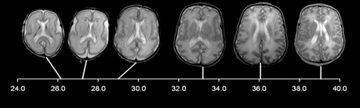
Autism’s origins: MRI scans of a developing human brain.
“We are very interested in studying how the brain develops normally and, by that means, to [get a reference point from which to better detect and study] abnormal development,” says Jo Hajnal, an imaging specialist at King’s College London and one of the leaders of the Developing Human Connectome Project.
Advertisement
The project is one of several efforts to create a three-dimensional map of the neuronal connections in the human brain. The U.S. BRAIN Initiative seeks to reconstruct the activity of every neuron in a brain (see “Why Obama’s Brain-Mapping Project Matters”) and the E.U. Human Brain Project seeks to create a detailed computational simulation of the human brain. The Allen Institute for Brain Science in Seattle also develops three-dimensional maps that combine gene activity data with structural detail of human and other animal brains and has recently described its own project to study the developing human brain by examining the cellular structure and organization of gene activity in post-mortem fetal brains.
This story is only available to subscribers.
Don’t settle for half the story.
Get paywall-free access to technology news for the here and now.
But the London group, which will use magnetic resonance imaging, or MRI, is different because it will examine the brains of growing fetuses. The project will be the first to produce a map of structural connections in the living human brain from the third trimester to the first weeks after birth. By combining different methods of MRI, the team will be able to determine the architecture of the brain down to a millimeter scale and overlay it with imaging that shows changes in blood flow in the brain that indicate neuron activity.
Mapping the prenatal brain is important because it will reveal when critical events are happening, says Ed Lein, a researcher at the Allen Institute who led the institute’s developing human brain atlas project. “And it’s important to know when events are happening because it’s also when they tend to go wrong,” he says.
A recent study on the brains of children who had died found that the majority of children with autism had disorganized patches of neurons in the cortex, the outer layer of the brain, which develops during the second and third trimesters. Another recent study, co-authored by Lein and based on the Allen Institute’s developing brain atlas, found that genes associated with autism tend to be active in the same outer layer of the brain.
The London group leading the Developing Human Connectome Project, which is expected to take six years, plans to scan 500 fetuses in the third trimester of pregnancy as well as 1,000 infants just days after birth. Some of those infants will be recruited because they have a close relative with autism. For the most part, children must be at least two years old to be diagnosed with autism, so the team will have to wait for years before they can go back and compare the brain scans of children with autism to those without the condition.
Before the Connectome project was launched, Hajnal and colleagues had already been working on techniques that would allow MRI to be safely used on fetuses. “It’s a completely safe thing to do, but you have to behave responsibly,” says Hajnal. Another challenge was movement—MRI generally requires the subject to coӧperate and stay still. So Hajnal and colleagues developed computational approaches that align the set of images collected during an MRI session into a coherent three-dimensional depiction of a brain, even if the fetus moved during the scan.