One of the largest connectomes published to date reveals how brains can detect motion.
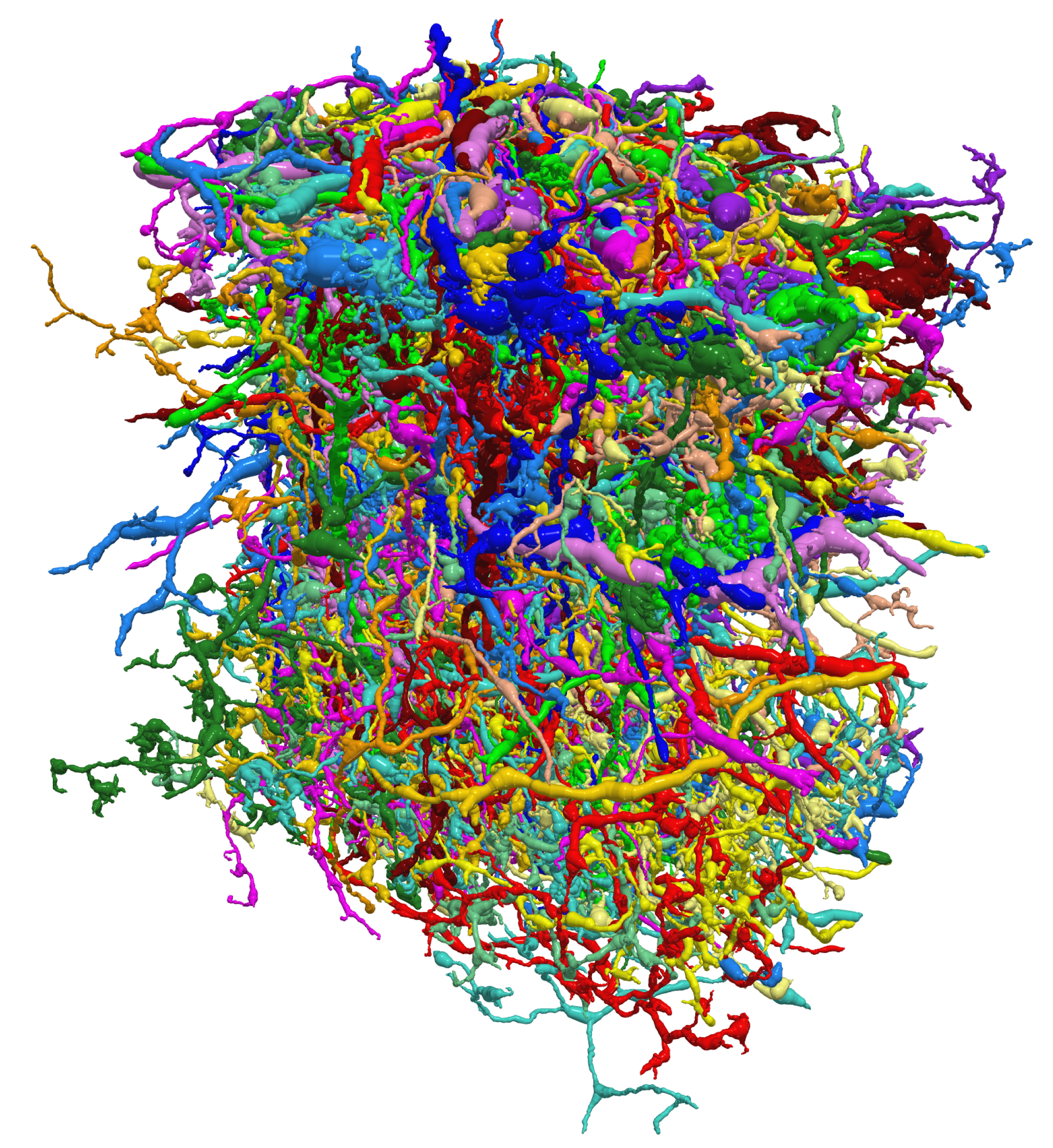
Flyswatter spotter: A reconstruction of 379 neurons involved in motion detection in the fruit fly.
Researchers at the Howard Hughes Institute’s Janelia Farm Research campus and their collaborators report in Nature on Wednesday that they were able to reconstruct the shapes and interconnections of neurons within a small part of the fly brain that is responsible for detecting visual motion.
Advertisement
By mapping the brain structure in such detail, the researchers gained new insight into how the brain detects movement. Their work is the latest example of many ongoing efforts in neuroscience to understand how the brain functions by building intricate diagrams of neuronal connections, or connectomes (see “Connectomics”).
This story is only available to subscribers.
Don’t settle for half the story.
Get paywall-free access to technology news for the here and now.
A theory for how neurons might work together to interpret motion had been around for about 60 years, but scientists didn’t know how the behavior was carried out by neuronal circuits, says senior author Dmitri Chklovskii. In part, that’s because tracing a neuronal circuit, even in the small brain of a fruit fly, is extremely difficult, he says.
To build their detailed three-dimensional map, Chklovskii and colleagues took pictures of very thin slices carved from a frozen fly brain and then stitched together more than 20,000 of those images. They were able to automate much of this reconstruction, but humans had to go through to check for errors. In total, around 14,400 person-hours were needed to build the connectome of 379 cells with 8,637 synaptic connections.
The researchers identified cells connected to each other in circuits that fit with the existing model for how brains detect motion. But to fully connect the structural information to behavior, patterns of neuron activity need to be combined with these detailed maps. Mapping out neuron activity in the brain is one of the major goals of the large scale neuroscience initiative announced by President Obama earlier this year (see “The Brain Activity Map”)
Along those lines, a more complete story of how the brain computes motion emerged when the wiring diagram was combined with the results of another study published in the same issue of Nature. Using fluorescent molecules that glow when neurons are active, a second team of scientists demonstrated that four subsets of neurons in the motion connectome each respond to motion in one of four cardinal directions: left, right, up, and down.
“With the combination of our anatomical work with theory, and the physiology and behavioral work of other labs, the whole story is starting to become clear now,” says Chklovskii. The connectomes of fly brains and human brains differ from one another, but “in both cases, the brains have to perform similar computations,” Chklovskii says. “The lessons learned will provide insight into solving how more complex computations are performed in the brains of animals, including vertebrates like us.”