Although tumor metastasis causes about 90 percent of cancer deaths, the mechanism that allows cancer cells to spread from one part of the body to another is not well understood. One key question is how they detach from the structural elements that normally hold tissues in place and then reattach themselves in a new site. A study from MIT researchers reveals some of the cellular adhesion molecules that are critical to this process, offering potential new drug targets.
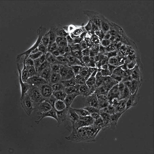
In this microscopic image, cancer cells adhere to a spot coated with molecules found in the extracellular matrix.
Cells inside the human body are usually tethered to a structural support system known as the extracellular matrix. Proteins called integrins, located on cell surfaces, form the anchors that attach the cells to the matrix. When cancer cells metastasize, these anchors let go.
Advertisement
In the new study, a group led by Sangeeta Bhatia, a professor of health sciences and technology and of electrical engineering and computer science, studied mice genetically engineered to develop lung cancer and compared the adhesion properties of cells from four types of tumors: primary lung tumors that later metastasized, primary lung tumors that did not metastasize, metastatic tumors that migrated from the lungs to nearby lymph nodes, and metastatic tumors that traveled to more distant locations such as the liver.
This story is only available to subscribers.
Don’t settle for half the story.
Get paywall-free access to technology news for the here and now.
The researchers developed technology that allowed them to expose each type of cell to about 800 different pairs of molecules found in the extracellular matrix and then measured how well the cells bound to the protein pairs. They were surprised to find that metastatic cells from different primary tumors were much more similar to each other in this respect than they were to the primary tumor from which they originally came. One pair of extracellular-matrix molecules that metastatic tumors stuck to especially well was fibronectin and galectin-3, both of which are made of proteins that contain or bind to sugars.
Previous studies have suggested that tumors pave the way for metastasis by secreting molecules that promote the development of environments hospitable to new cancer growth. Accumulation of galectin-3 and other molecules that help tumor cells colonize new sites may be part of this process, the researchers say.
The findings offer potential new ways to block metastasis by focusing on a specific cellular interaction rather than a particular gene mutation, says Nathan Reticker-Flynn, SM ’08, an MIT grad student and lead author of the study. “If those changes do confer a lot of metastatic potential, we can start thinking about how you target that interaction specifically,” he says.