MIT Technology Review Subscribe

The Brain Unveiled

A new imaging method offers a spectacular view of neural structures.
This image, generated from a living human brain, shows a subset of fibers. The red fibers in the middle and lower left are part of the corpus callosum, which connects the two halves of the brain.
Mapping Diffusion

Neural fibers in the brain are too tiny to image directly, so scientists map them by measuring the diffusion of water molecules along their length. The scientists first break the MRI image into “voxels,” or three-dimensional pixels, and calculate the speed at which water is moving through each voxel in every direction. Those data are represented here as peanut-shaped blobs. From each shape, the researchers can infer the most likely path of the various nerve fibers (red and blue lines) passing through that spot.
Another image of the peanut-shaped blobs that represent the diffusion of water molecules along neural fibers in the brain. From each shape, the researchers can infer the most likely path of the various nerve fibers (red and blue lines) passing through that spot.
The result is a detailed diagram like that of the brain stem shown here.
Emotion Control

To study specific circuits in the brain, scientists can isolate a subset of fibers. The circuit shown here represents the core of the human limbic system, which plays a central role in emotion and memory. The thick green bundle enclosed by the red circle is the cingulum bundle, which connects different parts of the cortex. The C-shaped blue fibers to the right, called the uncinate fasciculus, connect the temporal lobe, which regulates language and memory, with the frontal lobe, an area involved in higher executive function and planning. Damage to this circuit can result in the inability to form new memories and the loss of emotional control.
A Long Road

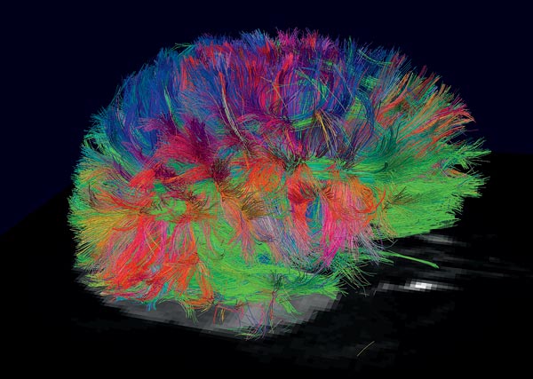
The complete brain of an owl monkey is shown here.
This is a subset of fibers from the brain of an owl monkey.
This image is the isolated optic tract, which relays visual signals from the eyes to the visual cortex, from the brain of an owl monkey. The blue lines at lower right represent nerve fibers connecting the eyes to the lateral geniculate nucleus (marked by the white ball), a pea-size ball of neurons that acts as a relay station for visual information. Those signals are then sent to the visual cortex, at the back of the head, via the blue and purple fibers that arc across the brain.
Advertisement
This story is only available to subscribers.

Don’t settle for half the story.
Get paywall-free access to technology news for the here and now.

Subscribe now Already a subscriber? Sign in
You’ve read all your free stories.

MIT Technology Review provides an intelligent and independent filter for the flood of information about technology.

Subscribe now Already a subscriber? Sign in
This is your last free story.
Sign in Subscribe now

Your daily newsletter about what’s up in emerging technology from MIT Technology Review.

Please, enter a valid email.
Privacy Policy
Submitting...
There was an error submitting the request.
Thanks for signing up!

Our most popular stories

Advertisement