In physicist Michael Feld’s MIT lab, researchers watch red blood cells vibrating and undulating in real time, thanks to a technology known as quantitative phase imaging. The technology splits a light wave in two, passes one wave through a cell, and then recombines it with the other wave.
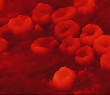

Analyzing the resulting interference pattern provides a remarkable view of living, moving cells not possible with electron microscopy, which requires careful sample preparation. Researchers in Feld’s lab are studying the dynamics of red blood cells’ membranes to gain insight into diseases such as malaria, leukemia, and sickle-cell anemia. Others are studying neuron dynamics. And while the MIT group has produced images with an astonishing 0.2-nanometer resolution, Feld ultimately hopes to create 3-D images of the inner structures of living cells, too.
Don’t settle for half the story.
Get paywall-free access to technology news for the here and now.
