Diffusion Tensor Imaging
Flipping through a pile of brain scans, a neurologist or psychiatrist would be hard pressed to pick out the one that belonged to a schizophrenic. Although schizophrenics suffer from profound mental problems – hallucinated conversations and imagined conspiracies are the best known – their brains look more or less normal.
This contradiction fascinated Kelvin Lim, a neuroscientist and psychiatrist at the University of Minnesota Medical School, when he began using imaging techniques such as magnetic resonance imaging (MRI) to study the schizophrenic brain in the early 1990s. Lim found subtle hints of brain structures gone awry, but to understand how these problems led to the strange symptoms of schizophrenia, he needed a closer look at the patients’ neuroanatomy than standard scans could provide.
Then, in 1996, a colleague told him about diffusion tensor imaging (DTI), a newly developed variation of MRI that allowed scientists to study the connections between different brain areas for the first time.
Diffusion Tensor Imaging
Key players
Peter Basser – Development of higher-resolution diffusion imaging techniques at National Institute of Child Health and Human Development; Aaron Field – Neurosurgery planning at University of Wisconsin-Madison; Michael Moseley – Assessment and early treatment of stroke at Stanford University
Lim has pioneered the use of DTI to understand psychiatric disease. He was one of the first to use the technology to uncover minute structural aberrations in the brains of schizophrenics. His group has recently found that memory and cognitive problems associated with schizophrenia, major but undertreated aspects of the disease, are linked to flaws in nerve fibers near the hippocampus, a brain area crucial for learning and memory. “DTI allows us to examine the brain in ways we hadn’t been able to before,” says Lim.
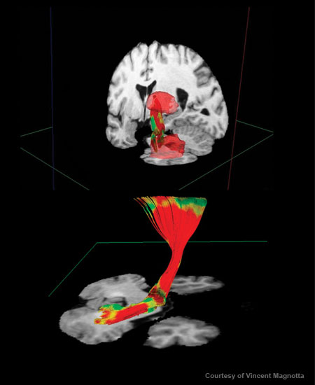
Conventional imaging techniques, such as structural MRI, reveal major anatomical features of the brain – gray matter, which is made up of nerve cell bodies. But neuroscientists believe that some diseases may be rooted in subtle “wiring” problems involving axons, the long, thin tails of neurons that carry electrical signals and constitute the brain’s white matter. With DTI, researchers can, for the first time, look at the complex network of nerve fibers connecting the different brain areas. Lim and his colleagues hope this sharper view of the brain will help better define neurological and psychiatric diseases and yield more-targeted treatments.
In DTI, radiologists use specific radio-frequency and magnetic field-gradient pulses to track the movement of water molecules in the brain. In most brain tissue, water molecules diffuse in all different directions. But they tend to diffuse along the length of axons, whose coating of white, fatty myelin holds them in. Scientists can create pictures of axons by analyzing the direction of water diffusion.
Following Lim’s lead, other neuroscientists have begun using DTI to study a host of disorders, including addiction, epilepsy, traumatic brain injury, and various neurodegenerative diseases. For instance, DTI studies have shown that chronic alcoholism degrades the white-matter connections in the brain, which may explain the cognitive problems seen in heavy drinkers. Other DTI projects are examining how the neurological scars left by stroke, multiple sclerosis, and amyotrophic lateral sclerosis (better known as Lou Gehrig’s disease) are linked to patients’ disabilities.
Lim is pushing the technology even further by combining it with findings from other fields, such as genetics, to unravel the mysteries of neurological and psychiatric disorders. Lim’s group has found, for instance, that healthy people with a genetic risk for developing Alzheimer’s disease have tiny structural defects in specific parts of the brain that are not shared by noncarriers. How these defects might be linked to the neurological problems of Alzheimer’s isn’t clear, but the researchers are trying to find the connection.
Lim and others also continue to refine DTI itself, striving for an even closer look at the brain’s microarchitecture. For example, current DTI techniques can easily image brain areas with large bundles of fibers all moving in the same direction, such as the corpus callosum, which connects the two hemispheres of the brain. But it has difficulty with areas such as the one where fibers leave the corpus callosum for other parts of the brain, which is a tangle of wires.
Researchers hope tools for studying white matter, like DTI, will help illuminate the mysteries of both healthy and diseased brains. Lim believes his own research into diseases like schizophrenia and Alzheimer’s could yield better diagnostics within 10 to 20 years – providing new hope for the next generation of patients.
Keep Reading
Most Popular
Large language models can do jaw-dropping things. But nobody knows exactly why.
And that's a problem. Figuring it out is one of the biggest scientific puzzles of our time and a crucial step towards controlling more powerful future models.
How scientists traced a mysterious covid case back to six toilets
When wastewater surveillance turns into a hunt for a single infected individual, the ethics get tricky.
The problem with plug-in hybrids? Their drivers.
Plug-in hybrids are often sold as a transition to EVs, but new data from Europe shows we’re still underestimating the emissions they produce.
Stay connected
Get the latest updates from
MIT Technology Review
Discover special offers, top stories, upcoming events, and more.