Printing Muscle
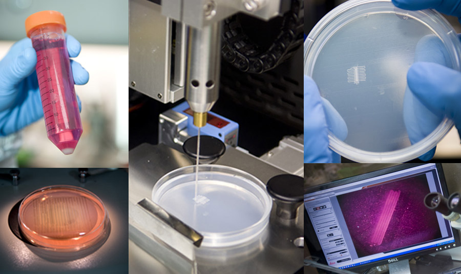
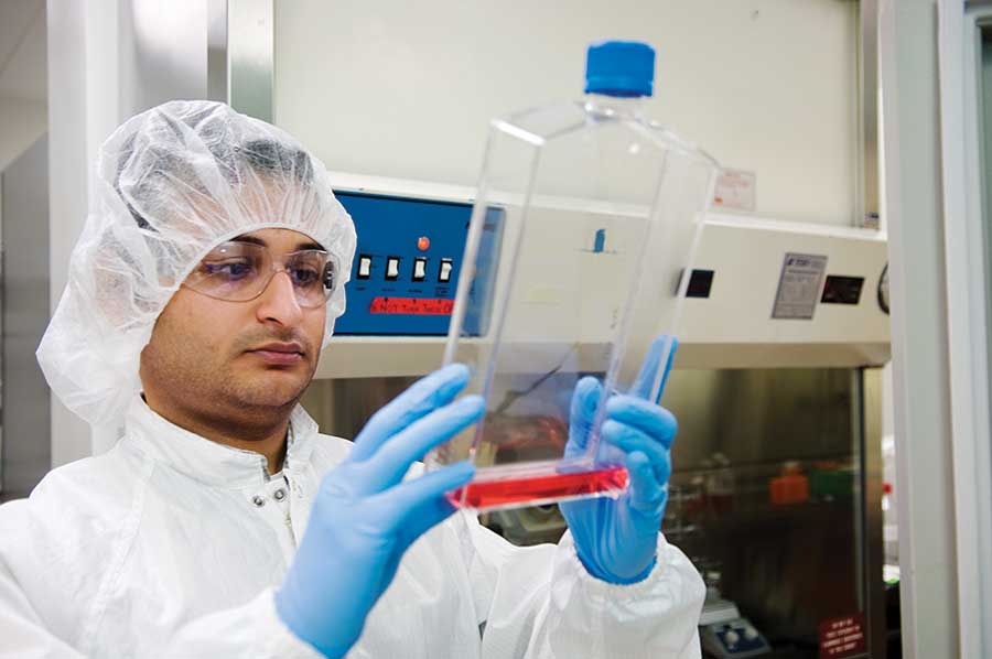
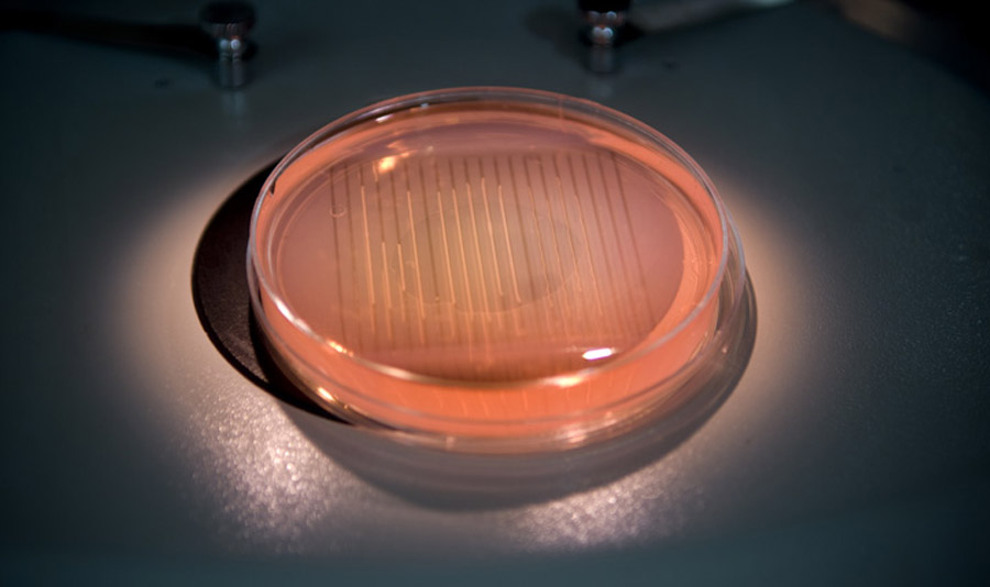
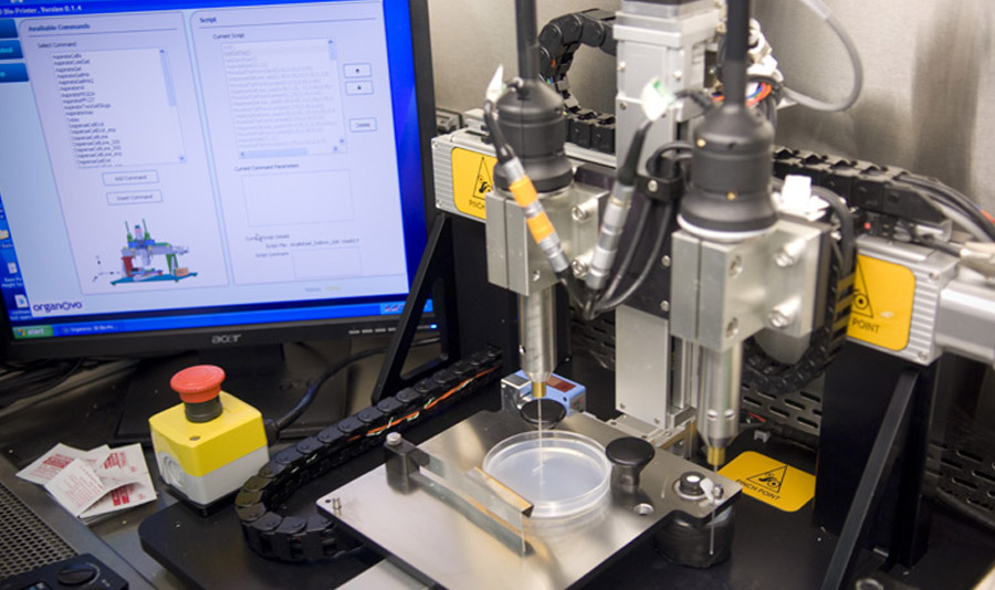
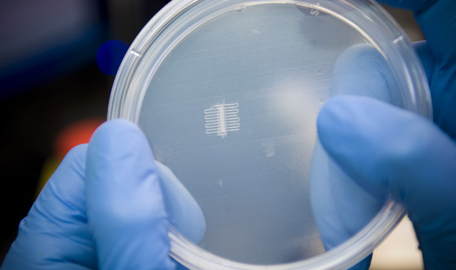
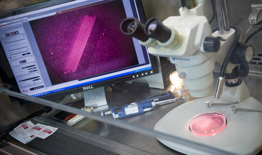
In a small clean room tucked into the back of San Diego–based startup Organovo, Chirag Khatiwala is building a thin layer of human skeletal muscle. He inserts a cartridge of specially prepared muscle cells into a 3-D printer, which then deposits them in uniform, closely spaced lines in a petri dish. This arrangement allows the cells to grow and interact until they form working muscle tissue that is nearly indistinguishable from something removed from a human subject.
The technology could fill a critical need. Many potential drugs that seem promising when tested in cell cultures or animals fail in clinical trials because cultures and animals are very different from human tissue. Because Organovo’s product is so similar to human tissue, it could help researchers identify drugs that will fail long before they reach clinical trials, potentially saving drug companies billions of dollars. So far, Organovo has built tissue of several types, including cardiac muscle, lung, and blood vessels.
Unlike some experimental approaches that have used ink-jet printers to deposit cells, Organovo’s technology enables cells to interact with each other much the way they do in the body. They are packed tightly together and incubated, prompting them to adhere to each other and trade chemical signals. When they’re printed, the cells are kept bunched together in a paste that helps them grow, migrate, and align themselves properly. Muscle cells, for example, orient themselves in the same direction to create tissue that can contract.
So far, Organovo has made only small pieces of tissue, but its ultimate goal is to use its 3-D printer to make complete organs for transplants. Because the organs would be printed from a patient’s own cells, there would be less danger of rejection.
Organovo plans to fund its organ-printing research with revenue from printing tissues to aid in drug development. The company is undertaking experiments to prove that its technology can help researchers detect drug toxicity earlier than is possible with other tests, and it is setting up partnerships with major companies, starting with the drug giant Pfizer.

Keep Reading
Most Popular
Large language models can do jaw-dropping things. But nobody knows exactly why.
And that's a problem. Figuring it out is one of the biggest scientific puzzles of our time and a crucial step towards controlling more powerful future models.
How scientists traced a mysterious covid case back to six toilets
When wastewater surveillance turns into a hunt for a single infected individual, the ethics get tricky.
The problem with plug-in hybrids? Their drivers.
Plug-in hybrids are often sold as a transition to EVs, but new data from Europe shows we’re still underestimating the emissions they produce.
Google DeepMind’s new generative model makes Super Mario–like games from scratch
Genie learns how to control games by watching hours and hours of video. It could help train next-gen robots too.
Stay connected
Get the latest updates from
MIT Technology Review
Discover special offers, top stories, upcoming events, and more.