Eye on Cells
In physicist Michael Feld’s MIT lab, researchers watch red blood cells vibrating and undulating in real time, thanks to a technology known as quantitative phase imaging. The technology splits a light wave in two, passes one wave through a cell, and then recombines it with the other wave.
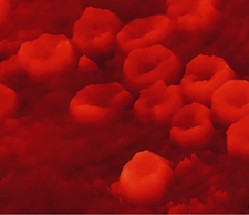
Analyzing the resulting interference pattern provides a remarkable view of living, moving cells not possible with electron microscopy, which requires careful sample preparation. Researchers in Feld’s lab are studying the dynamics of red blood cells’ membranes to gain insight into diseases such as malaria, leukemia, and sickle-cell anemia. Others are studying neuron dynamics. And while the MIT group has produced images with an astonishing 0.2-nanometer resolution, Feld ultimately hopes to create 3-D images of the inner structures of living cells, too.
Keep Reading
Most Popular
Large language models can do jaw-dropping things. But nobody knows exactly why.
And that's a problem. Figuring it out is one of the biggest scientific puzzles of our time and a crucial step towards controlling more powerful future models.
The problem with plug-in hybrids? Their drivers.
Plug-in hybrids are often sold as a transition to EVs, but new data from Europe shows we’re still underestimating the emissions they produce.
Google DeepMind’s new generative model makes Super Mario–like games from scratch
Genie learns how to control games by watching hours and hours of video. It could help train next-gen robots too.
How scientists traced a mysterious covid case back to six toilets
When wastewater surveillance turns into a hunt for a single infected individual, the ethics get tricky.
Stay connected
Get the latest updates from
MIT Technology Review
Discover special offers, top stories, upcoming events, and more.